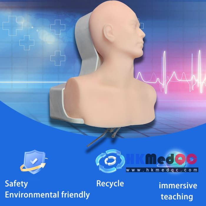
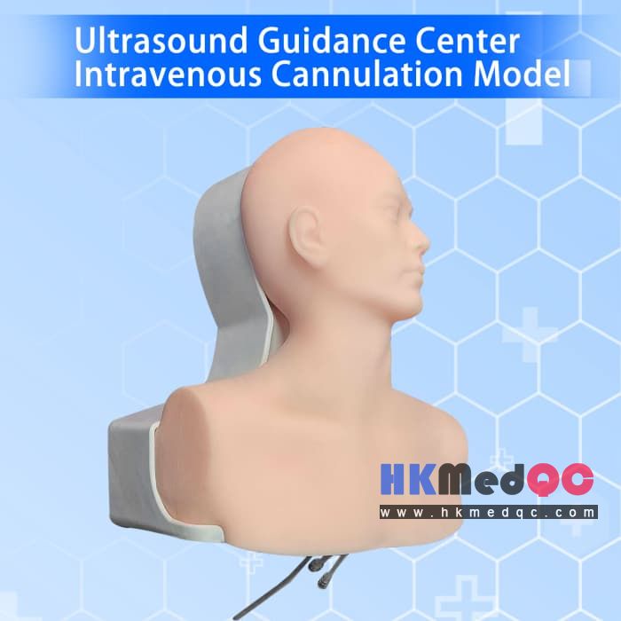
Center Intravenous Cannulation simulation ,Ultrasound Head-neck Puncture Model
Mode:HMQ-GVCM
Type:QA-Phantom
Contact: ken@hkmedqc.com

This high-fidelity ultrasound-compatible phantom is designed for hands-on training of central venous catheter placement procedures.
It simulates human tissue and vascular anatomy to deliver realistic ultrasound imaging and tactile feedback, allowing learners to practice ultrasound-guided cannulation safely and repeatedly without risk to live patients.
1.Product Highlights
Unmatched Realism for Authentic Training
- High-fidelity multi-layer phantom replicates human skin, subcutaneous tissue, and vascular anatomy for realistic tactile feedback and ultrasound imaging.
- Echogenic vessels clearly visible under ultrasound, with compressible veins and optional pulsatile arterial simulation for accurate vessel identification.
Comprehensive CVC Skill Training
- Supports full procedural training: needle insertion, guidewire manipulation, catheter threading, and confirmation of placement.
- Includes training for multiple access sites (IJ, subclavian, femoral) to build versatility in learners.
Risk-Free, Cost-Effective Education
- Eliminates the risks of practicing on live patients, making it ideal for medical students, residents, nurses, and allied health professionals.
- Durable, reusable construction reduces long-term training costs compared to single-use alternatives.
Efficient Skill Mastery
- Portable design enables both individual practice and group training sessions.
- Includes a step-by-step training guide with common ultrasound views and troubleshooting tips to accelerate learning.
Easy Maintenance & Compatibility
- Non-toxic, latex-free materials safe for repeated use and easy disinfection.
- Compatible with all major ultrasound gel types and standard clinical disinfectants.
2.Features and Applications:
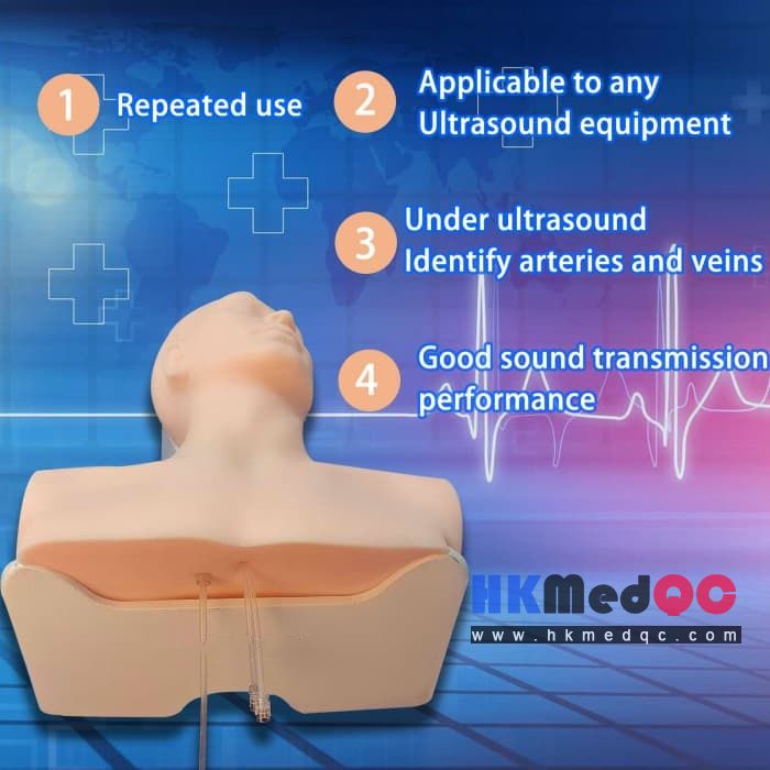
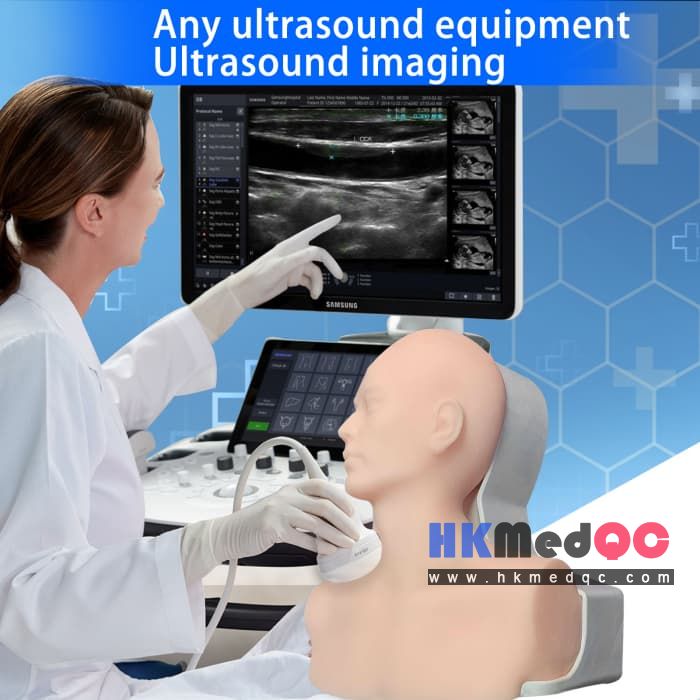
1. The product consists of a human body model, a stent, and a vascular pipeline device. Any ultrasound device can perform ultrasound imaging, and the image is close to the real human body image: using polymer materials equivalent to human tissue, ultrasound imaging is equivalent to human tissue density, subcutaneous blood vessel ultrasound detection, clear imaging of blood vessels, displaying low echo images, imaging guidance for blood vessel puncture, ultrasound simulation scanning direction, blood vessel depth position is equivalent to the actual human body, high and low echo imaging difference is uniform, blood pressure is present in the blood vessels, and the blood pressure of the internal jugular vein is simulated in reality. The puncture is used for blood return training.
2. Realistic imitation of a complete human head and neck with slight left rotation, exposing the neck operating area. The visible position of the clavicle is clear, and there is a hard bone sensation when touched. The transverse section of the internal jugular vein ultrasound image shows a low echo elliptical shape, approximately 1x1.3cm, closely adjacent to the carotid artery. The vascular ultrasound imaging shows a low echo circular shape with a diameter of about 6mm. The position relationship between the high-frequency ultrasound probe's transverse and longitudinal sections of the blood vessels is clear.
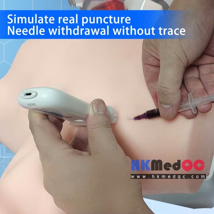
3. Arterial blood vessels are filled with bright red fluid, venous blood vessels are filled with blue fluid, and external venous blood pressure is applied to the internal jugular vein. After successful puncture, there is a blood return function. If there is an operational error during the operation, the liquid color displayed on the syringe can quickly identify arterial blood or venous blood to determine whether the puncture is successful.
4. Can be reused multiple times, and the material is repairable.
5. Good sound transmission performance, capable of displaying different grayscale sound wave images in different tissues
3.Target Users & Applications
- Medical schools and residency programs
- Nursing and allied health training centers
- Hospital simulation labs and continuing medical education (CME) programs
- Critical care, emergency medicine, and anesthesia departments
- Medical simulation centers and skills labs worldwide
4.Usage & Maintenance Notes
- Clean phantom surface with mild disinfectant after each use; remove all ultrasound gel residue.
- Avoid sharp objects or excessive force that may damage the self-healing tissue.
- Store in a cool, dry place away from direct sunlight to preserve material integrity.
- Replaceable vessel inserts (optional upgrade) available for extended product lifespan.
SAG: Ultrasound-guided CVC training model,Central venous catheter phantom,Ultrasound vascular access trainer,Internal jugular vein cannulation model