KS215T-1 Tissue-Mimicking Ultrasound Elastography Phantom, KS215T-1 Multi-Target
Mode:KS215T-1
Type:QA-Phantom
Contact: ken@hkmedqc.com
KS215T-1 Tissue-Mimicking Ultrasound Elastography Phantom, KS215T-1 Multi-Target.PDF

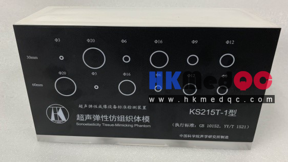
The composition and structure of the KS215T-1 series of ultrasonic elastic tissue-like phantom models are as follows:
(1) The entire series consists of 4 independent phantom models with the same background material but different targets;
(2) The side and bottom plates of the phantom models are both 10mm thick organic glass, and the top surface is covered with a composite film sound window;
(3) Two circular holes with a diameter of 36mm are opened on the bottom plate, and the openings are sealed with high-quality rubber for liquid injection maintenance;
(4) The size of the background material is: 210mm in length, 60mm in width, and 110mm in height (depth);
(5) The target is cylindrical, with 6 diameters, namely Φ3, Φ6, Φ9, Φ12, Φ16, and Φ20mm;
(6) The targets are arranged in two rows, with the axis located 30mm and 60mm below the sound window respectively, and the horizontal interval is uniformly 30mm. The starting center point of the target is 40mm away from the edge of the phantom model horizontally;
(7) The axis of the cylinder is along the 60mm direction of the background material, perpendicular to the front and rear shell plates, and parallel to the surface of the sound window, and they are parallel to each other.
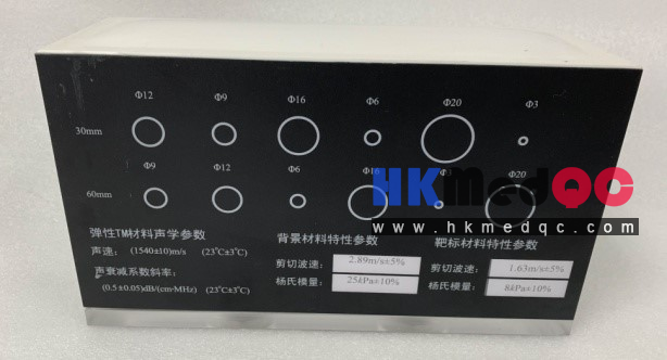
Acoustic-mechanical properties of the background and target materials
(1) At 23℃, the longitudinal wave speed of the background and target materials is (1540±10)m/s;
(2) At 23℃, the slope of the longitudinal wave attenuation coefficient of the background and target materials is (0.5±0.05)dB/(cm MHz);
(3) The background materials of all the models in the series are the same at 23℃, with a shear wave velocity of 2.89m/s±5% and a Young's modulus of 25kPa±10%;
(4) The shear wave velocities of the target materials of each model in the series are 1.63m/s±5%, 2.16m/s±5%, 3.87m/s±5%, 5.16m/s±5%, respectively, and the Young's moduli are 8kPa±10%, 14kPa±10%, 45kPa±10%, 80kPa±10%, respectively.
Product Details:
(1) The entire series consists of 4 independent phantom models with the same background material but different targets;
(2) The side and bottom plates of the phantom models are both 10mm thick organic glass, and the top surface is covered with a composite film sound window;
(3) Two circular holes with a diameter of 36mm are opened on the bottom plate, and the openings are sealed with high-quality rubber for liquid injection maintenance;
(4) The size of the background material is: 210mm in length, 60mm in width, and 110mm in height (depth);
(5) The target is cylindrical, with 6 diameters, namely Φ3, Φ6, Φ9, Φ12, Φ16, and Φ20mm;
(6) The targets are arranged in two rows, with the axis located 30mm and 60mm below the sound window respectively, and the horizontal interval is uniformly 30mm. The starting center point of the target is 40mm away from the edge of the phantom model horizontally;
(7) The axis of the cylinder is along the 60mm direction of the background material, perpendicular to the front and rear shell plates, and parallel to the surface of the sound window, and they are parallel to each other.
Acoustic-mechanical properties of the background and target materials
(1) At 23℃, the longitudinal wave speed of the background and target materials is (1540±10)m/s;
(2) At 23℃, the slope of the longitudinal wave attenuation coefficient of the background and target materials is (0.5±0.05)dB/(cm MHz);
(3) The background materials of all the models in the series are the same at 23℃, with a shear wave velocity of 2.89m/s±5% and a Young's modulus of 25kPa±10%;
(4) The shear wave velocities of the target materials of each model in the series are 1.63m/s±5%, 2.16m/s±5%, 3.87m/s±5%, 5.16m/s±5%, respectively, and the Young's moduli are 8kPa±10%, 14kPa±10%, 45kPa±10%, 80kPa±10%, respectively.
Classification and basic characteristics of ultrasound elastography image formation equipment
| Method Category | Force Application Type | Force Application Method | Display Characteristic | Qualitative/Quantitative | Imaging/Measurement Mode | |
| Displacement or Strain Imaging | Strain Elastography | Quasi-static Force | Applied externally through the skin using a mechanical device | Strain | Qualitative | Full-area imaging, updated at diagnostic ultrasound frame rate |
| Strain Rate Imaging | Quasi-static Force | Applied internally via physiological processes | Strain Rate | Qualitative | Full-area imaging, updated at diagnostic ultrasound frame rate | |
| Acoustic Radiation Force Impulse (ARFI) Imaging | Dynamic force | Applied at a specified depth using focused radiation force pulses generated by ultrasound | Displacement | Qualitative | Single-frame imaging within the sampling box | |
| Shear Wave Speed Measurement | Transient Elastography | Dynamic force | Applied externally through the skin using a mechanical device | Shear Wave Speed | Quantitative | Single measurement, average value along the sound beam |
| Point Shear Wave Elastography (also called Acoustic Radiation Force Quantification, ARFQ) | Dynamic force | Applied at a specified depth using focused radiation force generated by ultrasound | Shear Wave Speed | Quantitative | Single measurement, average value within the region of interest | |
| Shear Wave Speed Imaging | Shear Wave Elastography | Dynamic force | Applied at several depths using focused radiation force generated by ultrasound | Shear Wave Speed | Quantitative | Single-frame imaging within the color box |
| Dynamic force | The focused radiation force generated by ultrasound sweeps through a range of depths faster than the shear wave speed, forming a Mach cone | Shear Wave Speed | Quantitative | Imaging within the color box, updated at a rate of several frames per second | ||
Product Introduction:
Used for testing the performance of quasi-static strain elastography system, acoustic radiation force impulse elastography system, point shear wave elastography system, and (ultrasonic velocity) shear wave elastography system.
Technical Specifications:
1. Basic Structure
The side and bottom plates of the body model shell are both made of 10mm thick organic glass, and the top surface is covered with a composite film sound window.
(2) Two circular holes with a diameter of 36mm are drilled on the base plate. The top is sealed with high-quality rubber, which can be used for filling and maintenance purposes.
(3) The dimensions of the background material are: 210mm in length, 60mm in width, and 110mm in height (depth).
(4) The target is cylindrical in shape, and its diameters are available in six different sizes: Ø3, Ø6, Ø9, Ø12, Ø16, and Ø20 mm.
(5) The target is divided into two rows, with the axis located 30mm and 60mm below the sound window respectively. The distance between each axis along the horizontal direction is uniformly 30mm.
(6) The axis of the cylinder is oriented in the 60mm direction of the background material, perpendicular to the front and rear shell plates, parallel to the surface of the sound window, and parallel to each other.
(7) The targets for each platform are as listed in Table 2 and shown in Figure 2, arranged crosswise according to their diameters.
| ←Left Right→ | ||||||
| Upper layer | Ø3 | Ø20 | Ø6 | Ø16 | Ø 9 | Ø12 |
| Lower layer | Ø20 | Ø3 | Ø16 | Ø 6 | Ø12 | Ø9 |
Product Details:
KS215T-1 Inclined Ultrasonic Elastography Tissue-like Model
Standard configuration: One integrated ultrasonic elasticity tissue-like phantom, one user manual, one test report, one certificate of conformity, one packing list, and one portable box.
SAG: KS215T-1 Ultrasound phantom,Elastography phantom,Tissue mimicking phantom,Multi-target phantom,Strain elasticity phantom,Medical calibration phantom,Ultrasound test phantom,Soft tissue phantom,Diagnostic ultrasound phantom,KS215T-1 phantom