
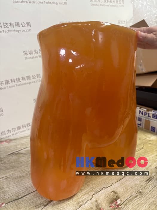
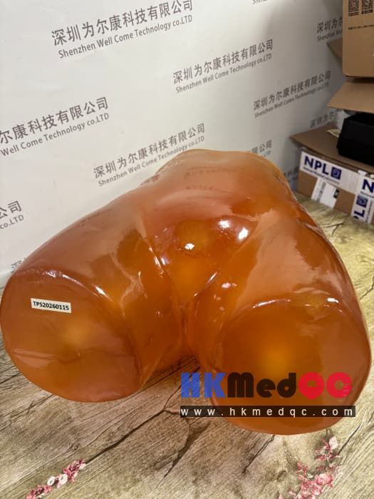
PE-A01 Adult Pelvis Model,Adult pelvic abdominal model
Mode:PE-A01
Type:QA-Phantom
Contact: ken@hkmedqc.com
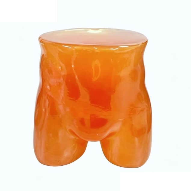
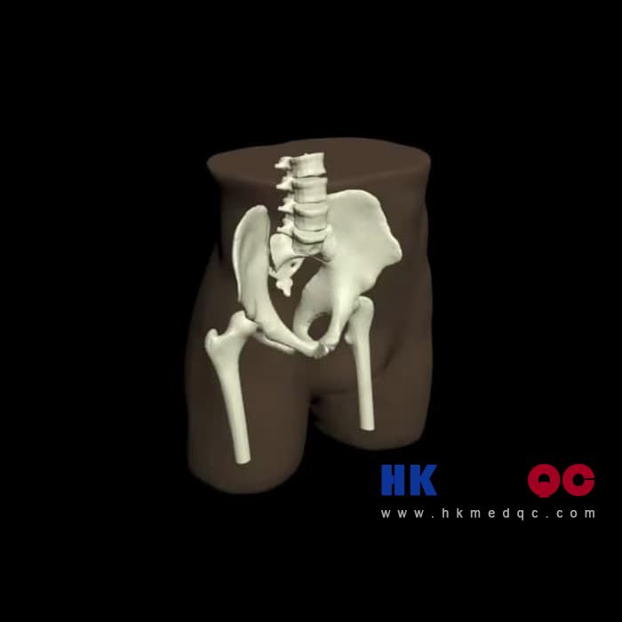
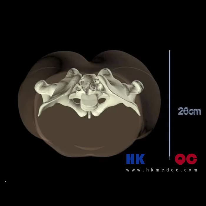
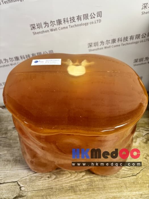
The original factory-produced human biomimetic model closely replicates the adult's lumbar vertebrae, sacrum, complete pelvic bones, as well as abdominal fat and muscle soft tissue anatomical structures. It is made of human tissue equivalent solid materials, and its X-ray attenuation, CT value, ultrasonic acoustic impedance are highly matched with real human bones and soft tissues, suitable for multi-modal imaging scans such as X-ray, CT, and B-ultrasound. It is mainly used for quality control and acceptance of radiological imaging equipment, medical staff practical training, medical school teaching, and imaging research experiments.
2.Universal three-modal compatibility: compatible with all mainstream imaging equipment such as X-ray photography, CT tomography, and abdominal pelvic B-ultrasound.
3.Solid and integrated one-piece design, without internal liquids, preventing leakage and aging, capable of repeated scanning for tens of thousands of times.
4.Simulates the human body's shape contour, conforms to the routine scanning positions of clinical patients, making training and practical operations more closely resemble real diagnosis and treatment.
5.Customizable: can adjust the ratio of soft tissues, add simulated lesions, and modify local anatomical structures as needed.
In terms of the application of magnetic resonance imaging, this simulated tissue has T2 relaxation times that are close to the actual values, making it the best choice for all T2-weighted magnetic resonance imaging methods. Using the proton density imaging method can also yield very good results. This simulated tissue can also be used for T1-weighted imaging, but the T1 value is slightly insufficient, approximately 100 milliseconds.
The skeletal model will adopt a realistic three-layer structure with pores inside, which can be adjusted according to the requirements of various projects. Moreover, this model can also be customized in size and shape based on an individual's CT scan data.
Practical exercises for pelvic and abdominal scanning techniques and positioning by resident doctors and ultrasound technicians
Imaging equipment manufacturers' image effect debugging during the research and development stage and factory quality inspection
Medical imaging majors in universities and research institutes, anatomical teaching, and subject scanning experiments
Compliance verification of imaging performance by third-party medical device testing institutions
PE-A01 Pelvic and Abdominal Model Host, original factory English manual, hard protective shockproof storage box, warranty card
Avoid scratches, heavy impacts or falls caused by sharp objects, and prevent surface damage;
Store at room temperature in a dark place, and avoid long-term exposure to high temperatures;
Only use neutral alcohol for cleaning; do not use strong acid or strong alkali disinfectants;
If a compatible version for MRI and pelvic model is needed, you can contact for customization selection.
Core advantage
1.Highly detailed anatomical structure: fully includes the lumbar vertebrae, sacrum, ilium, ischium, and pubis of the pelvis, along with the surrounding abdominal muscles and fatty soft tissues.2.Universal three-modal compatibility: compatible with all mainstream imaging equipment such as X-ray photography, CT tomography, and abdominal pelvic B-ultrasound.
3.Solid and integrated one-piece design, without internal liquids, preventing leakage and aging, capable of repeated scanning for tens of thousands of times.
4.Simulates the human body's shape contour, conforms to the routine scanning positions of clinical patients, making training and practical operations more closely resemble real diagnosis and treatment.
5.Customizable: can adjust the ratio of soft tissues, add simulated lesions, and modify local anatomical structures as needed.
In terms of the application of magnetic resonance imaging, this simulated tissue has T2 relaxation times that are close to the actual values, making it the best choice for all T2-weighted magnetic resonance imaging methods. Using the proton density imaging method can also yield very good results. This simulated tissue can also be used for T1-weighted imaging, but the T1 value is slightly insufficient, approximately 100 milliseconds.
The skeletal model will adopt a realistic three-layer structure with pores inside, which can be adjusted according to the requirements of various projects. Moreover, this model can also be customized in size and shape based on an individual's CT scan data.
Applicable scenarios
Annual inspection and quality control of imaging equipment in the hospital's radiology and ultrasound departments, acceptance of new equipment upon arrivalPractical exercises for pelvic and abdominal scanning techniques and positioning by resident doctors and ultrasound technicians
Imaging equipment manufacturers' image effect debugging during the research and development stage and factory quality inspection
Medical imaging majors in universities and research institutes, anatomical teaching, and subject scanning experiments
Compliance verification of imaging performance by third-party medical device testing institutions
Standardized list:
PE-A01 Pelvic and Abdominal Model Host, original factory English manual, hard protective shockproof storage box, warranty card
Usage Reminder
Avoid scratches, heavy impacts or falls caused by sharp objects, and prevent surface damage;Store at room temperature in a dark place, and avoid long-term exposure to high temperatures;
Only use neutral alcohol for cleaning; do not use strong acid or strong alkali disinfectants;
If a compatible version for MRI and pelvic model is needed, you can contact for customization selection.
SAG: PE-A01,Adult pelvic model,abdominal model,PE-A01 adult pelvic model,adult pelvic model,PE-A01 abdominal model,PE-A01 adult pelvic model,medical imaging QC phantom,adult pelvic phantom