
Cranial CT Phantom,CT/ CBCT imaging Head CT phantom
Mode:QRMCranial-CT
Type:QA-Phantom
Contact: ken@hkmedqc.com
Cranial CT Phantom,CT/ CBCT imaging Head CT phantom.PDF
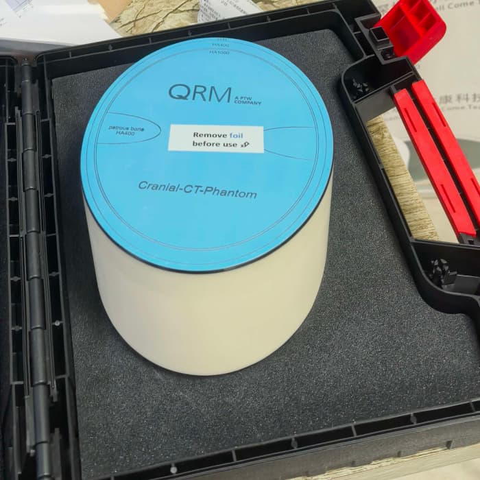
The cranial CT phantom is used for CT/CBCT imaging, simulating the human skull and brain anatomical structures.
1. Acceptance testing and regular quality control for head CT and oral CBCT equipment
2. Evaluation of low-contrast detection capability, spatial resolution, CT value accuracy, image noise, and uniformity
3. Medical physics research, image algorithm verification, and technician scanning training

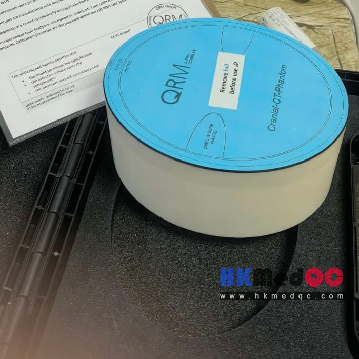
Simulating human skull and brain anatomical structures is used for CT imaging.
It is used to study artifacts such as low contrast, beam hardening, and partial volume effect.
The cranial CT phantom is based on brain tissue equivalent plastic and has a constant CT value under different kV settings. It is surrounded by a bone structure that simulates the X-ray absorption characteristics of the human skull.
The cranial CT phantom contains two different parts:
One part simulates the base of the skull and includes uniform brain tissue and bone structure equivalent to the temporal bone. This part is suitable for displaying and evaluating the image quality of the skull base region, which is particularly susceptible to beam hardening effect, partial volume effect, and artifacts caused by scanner instability (such as anode jitter).
The second part is used to test the low contrast resolution capability of the imaging system. Several low contrast structures are set in the uniform brain tissue to simulate the typical cerebral cortex structure and the low contrast characteristics of the brain center region.
Core Parameters
- Model: QRM-20113 Cranial CT Phantom
- Dimensions: 140 mm × 180 mm, height 88 mm
- Weight: 2.0 kg
- Main material: Brain tissue equivalent material (≈35HU)
- Built-in modules: Low-contrast lesion module, spatial resolution line pair module, uniformity test area
- Accessories: Hard protective carrying case
- Dimensions: 140 mm × 180 mm, height 88 mm
- Weight: 2.0 kg
- Main material: Brain tissue equivalent material (≈35HU)
- Built-in modules: Low-contrast lesion module, spatial resolution line pair module, uniformity test area
- Accessories: Hard protective carrying case
Application Scenarios
1. Acceptance testing and regular quality control for head CT and oral CBCT equipment
2. Evaluation of low-contrast detection capability, spatial resolution, CT value accuracy, image noise, and uniformity
3. Medical physics research, image algorithm verification, and technician scanning training
SAG: Cranial CT Phantom,QRM-20113,QRM phantom,medical head phantom,CBCT phantom,CT image quality control phantom,radiology phantom,x-ray test phantom,CT calibration phantom,medical phantom for radiology