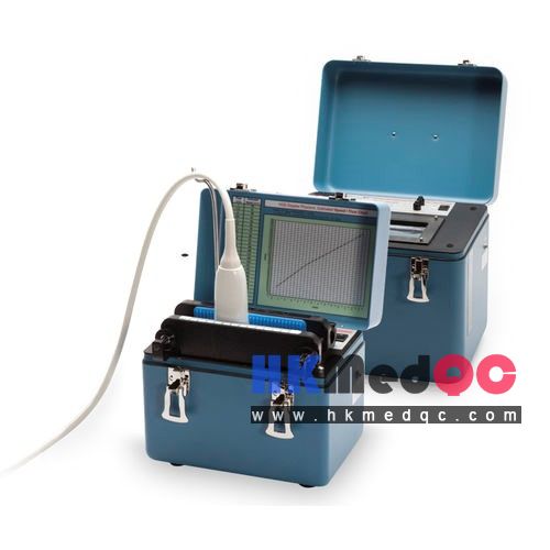
US-CT Simulated Liver Puncture Model,US-CT Multi-Modal Ultrasound Phantom
Mode:US-CT
Type:QA-Phantom
Contact: ken@hkmedqc.com
US-CT Simulated Liver Puncture Model,US-CT Multi-Modal Ultrasound Phantom.PDF

The Liver Puncture Phantom (US-CT Multi-Modal) is a custom-manufactured product containing tissue-mimicking materials. It is designed for use in training demonstrations for ultrasound-guided puncture procedures and is capable of generating images under both ultrasound and X-CT modalities.
Key Technical Specifications:
Model: Liver Puncture Simulator – US-CT Multi-Modal Ultrasound Phantom
Housing: Acrylic (Metal-free; no CT artifacts)
External Dimensions: 280 × 170 × 160 mm
Internal Dimensions: 240 × 150 × 140 mm
Weight: Approx. 10 kg
Acoustic Material: TM Human Tissue-Mimicking Gel
Speed of Sound: 1540 ± 10 m/s (at 23°C)
Acoustic Attenuation: ≤ 0.10 dB/cm/MHz
Acoustic Window: 70 μm Polyester Film, 140 × 150 mm
Modality Compatibility: Dual-Mode Imaging (Ultrasound [US] + X-CT)
Application: Training for Liver Puncture, Biopsy, and Ablation; Image Fusion Practice
Technical Specifications
Based on the national standard GB10152—2009 and tailored to the technical requirements for ultrasound phantoms in training applications, the technical parameters of this phantom are as follows:
Speed of Sound within Internal TM Material (Human Tissue-Mimicking Material): (1540 ± 10) m/s (at 23°C ± 3°C)
Slope of Background Acoustic Attenuation Coefficient: Not greater than 0.10 dB/cm/MHz (at 23°C ± 3°C)
Left Acoustic Window Material: 70 µm thick polyester film.
Left Acoustic Window Opening Dimensions: 140 mm × 150 mm.
The phantom features a cubic overall structure, with its four sides bonded together using acrylic (Plexiglas) panels; the entire assembly contains no metallic components. The structural layout is illustrated in Figure 2. The interior of the phantom consists of a background tissue-mimicking material composed of a transparent aqueous gel, which also encapsulates a semi-transparent aqueous gel structure shaped like a human liver. Embedded within this semi-transparent, liver-mimicking structure are several dark-colored cylindrical targets, distributed uniformly throughout the "liver" volume. The axes of these cylindrical targets run parallel to the thickness dimension of the phantom; these targets serve as reference points for puncture training. Under both ultrasound and X-CT imaging modes, these targets generate distinct image contrast relative to the surrounding tissue-mimicking background material.
The top surface of the phantom is covered by a 1.5 mm thick semi-transparent silicone rubber membrane, which is secured in place by the rim of an outer water-bath frame. This outer frame is constructed from plastic and is fastened to the edges of the phantom's housing via hand-tightened screws located on all four sides. The silicone rubber membrane functions as both the acoustic window and the puncture entry point, while also serving the purpose of retaining the internal aqueous gel fluid.
SAG: Liver Phantom,US-CT Dual-modal,Puncture Training,Ultrasound Phantom,CT Imaging Phantom,Tissue Mimicking,Interventional Training,Biopsy Simulation,Medical Calibration,Clinical Simulation