
PhantomX WEK51-04 CTA AVM Head Phantom,CT angiography head model
Mode:WEK51-04
Type:QA-Phantom
Contact: ken@hkmedqc.com
PhantomX WEK51-04 CTA AVM Head Phantom,CT angiography head model.PDF
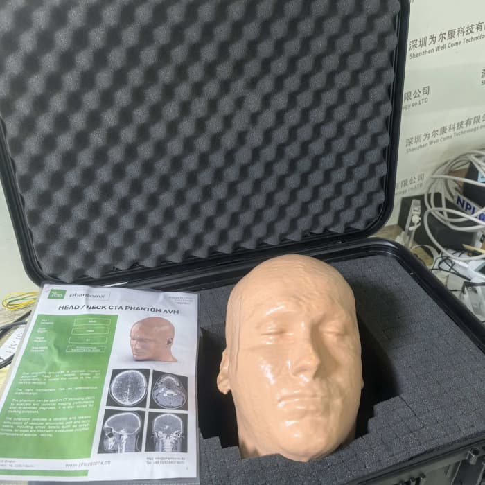
This model simulates the head (CT angiography scene) after contrast agent enhancement during the arterial phase, with the simulation range extending from the top of the skull to the fifth cervical vertebra.
On the right side of the model's cerebral hemisphere, an arteriovenous malformation lesion is set.
This model is suitable for CT equipment (including cone-beam CT) and can be used for evaluating, optimizing imaging performance, and implementing artificial intelligence-assisted diagnostic algorithms. It can also be used for clinical teaching and training.
The model is highly detailed and accurately reproduces vascular, soft tissue, and bone structures, including minute anatomical structures such as lymph nodes; it simulates gas cavities filled with cellulose polymer composite materials, corresponding to a CT value of approximately -160 Hounsfield Units (HU).
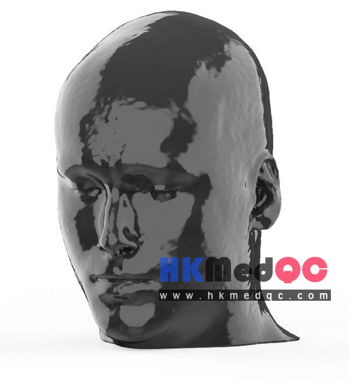
An arteriovenous malformation (AVM) lesion is located in the right cerebral hemisphere.
2.Neurointerventional department: Doctors' practice of AVM lesion localization, planning of interventional catheter paths, practical training for CTA image reading and analysis
3.CT imaging equipment manufacturer: Research and development of cerebral vascular imaging algorithms, adjustment of reconstruction sequences, performance testing of the entire machine before leaving the factory
4.Medical colleges, neuroimaging laboratories: Teaching of cerebral vascular diseases, research experiments on post-processing reconstruction of CTA images
5.Third-party medical imaging testing institutions: Compliance verification of CT angiography imaging performance
6.Contrast agent manufacturers: Comparison of cerebral vascular filling effects with different iodine contrast agents, testing of imaging development timeliness
2. The body model is coated with a protective layer. If the protective layer is undamaged, you can clean the body model with a damp cloth (water or a neutral cleaner).
3. Avoid direct sunlight.
4. Maintain the storage temperature between 10°C and 30°C. If the model is exposed to temperatures lower than 10°C or higher than 45°C, it may be severely damaged.
5. This model is not suitable for dose measurement using dosimeters, and it is also not appropriate for material characterization using dual-energy CT.
6. This model has not obtained the medical device certification.
7. The air voids are filled with a cellulose polymer composite material with a density of approximately -160 HU.
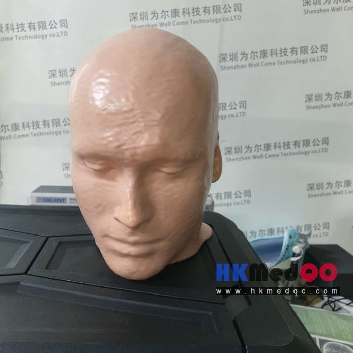
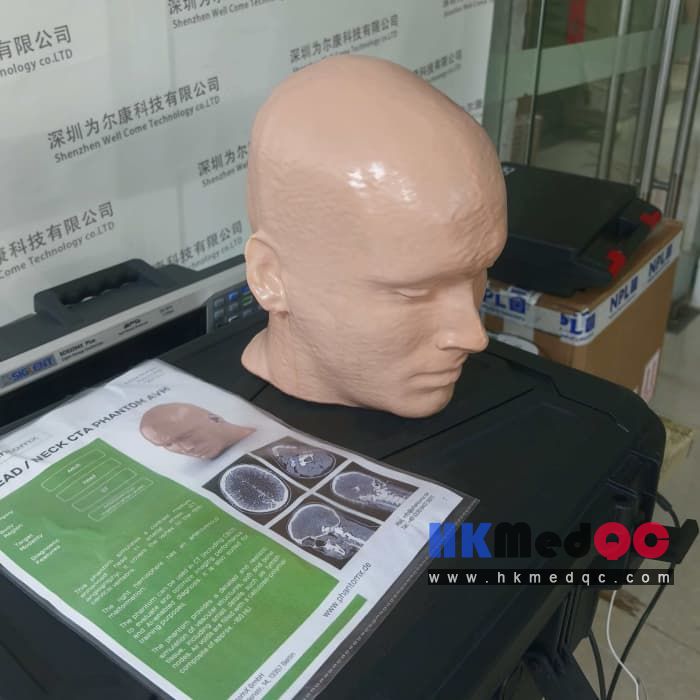
On the right side of the model's cerebral hemisphere, an arteriovenous malformation lesion is set.
This model is suitable for CT equipment (including cone-beam CT) and can be used for evaluating, optimizing imaging performance, and implementing artificial intelligence-assisted diagnostic algorithms. It can also be used for clinical teaching and training.
The model is highly detailed and accurately reproduces vascular, soft tissue, and bone structures, including minute anatomical structures such as lymph nodes; it simulates gas cavities filled with cellulose polymer composite materials, corresponding to a CT value of approximately -160 Hounsfield Units (HU).
| Item | Parameters |
| Overall Dimensions | Approx. 186 × 224 × 269 mm |
| Weight | Approx. 5120 g |
| Base Material | Cellulose polymer composite material |
| Optimal Tube Voltage | 120 kVp (see Page 3)Customizable and adjustable according to customer requirements |
Diagnostic Features
It can realistically simulate cranial blood vessels, bones and soft tissues of the human head.An arteriovenous malformation (AVM) lesion is located in the right cerebral hemisphere.
Suitable for all scenarios
1.Hospital radiology department: New machine acceptance for cranial CTA equipment, annual image quality control, adjustment of scan slice thickness / contrast sequence parameters2.Neurointerventional department: Doctors' practice of AVM lesion localization, planning of interventional catheter paths, practical training for CTA image reading and analysis
3.CT imaging equipment manufacturer: Research and development of cerebral vascular imaging algorithms, adjustment of reconstruction sequences, performance testing of the entire machine before leaving the factory
4.Medical colleges, neuroimaging laboratories: Teaching of cerebral vascular diseases, research experiments on post-processing reconstruction of CTA images
5.Third-party medical imaging testing institutions: Compliance verification of CT angiography imaging performance
6.Contrast agent manufacturers: Comparison of cerebral vascular filling effects with different iodine contrast agents, testing of imaging development timeliness
Adaptation situation:
1. This model is made of cellulose polymer composite material and has characteristics similar to hardwood. If handled carefully, it will last for a long time.2. The body model is coated with a protective layer. If the protective layer is undamaged, you can clean the body model with a damp cloth (water or a neutral cleaner).
3. Avoid direct sunlight.
4. Maintain the storage temperature between 10°C and 30°C. If the model is exposed to temperatures lower than 10°C or higher than 45°C, it may be severely damaged.
5. This model is not suitable for dose measurement using dosimeters, and it is also not appropriate for material characterization using dual-energy CT.
6. This model has not obtained the medical device certification.
7. The air voids are filled with a cellulose polymer composite material with a density of approximately -160 HU.
For more effects and DICOM data, please contact us.
WEK51-04 CTA AVM Head Mold, WEK51-04 CT Angiography Head Mold Product Physical Picture:
SAG: WEK51-04,WEK51-04 CT Angiography Head Model,CT Angiography Head Model,Angiography Head Model,CTA AVM Head Mold,CTA Head Mold,CTA AVM Head Model,CTA Head Model,PhantomX 51-04,WEK51-04 phantom,CTA head phantom