KS105FC-1 Puncture Training Model,KS105FC-1 Ultrasound Imaging Phantom
Mode:KS105FC-1
Type:QA-Phantom
Contact: ken@hkmedqc.com
KS105FC-1 Puncture Training Model,KS105FC-1 Ultrasound Imaging Phantom.PDF

KS105FC-1: Basic model, suitable for beginners for needle-punching practice.
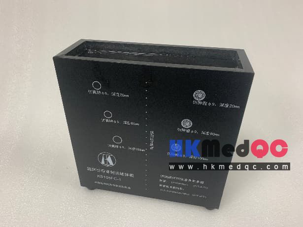
The puncture training mannequin set is a custom-made product. It is equipped with materials that simulate human tissues and is used for ultrasound imaging puncture training. The model is named KS105FC-1. It is filled with ultrasound-simulated human tissue materials. Inside the mannequin, there is a row of depth linear target lines arranged along the depth direction. On both sides of the target lines, cylindrical target markers such as tumors and cysts are buried. The depths of the simulated tumors are 30mm, 70mm, and 110mm, with a diameter of 9mm. The axes of the lesion columns are parallel to the target lines. The center depth of the 6mm-diameter simulated tumor is 30mm and 70mm, and the depth of the 9mm-diameter simulated cyst is 110mm. This mannequin is used to complement ultrasound-guided puncture training and demonstrations. It can be used for multiple punctures. The puncture holes can be closed after the puncture needle is removed, and there will be no puncture marks in the ultrasound image.
Basic Structure :
1. The outer shell is made of acrylic glass.
2. The overall dimensions are 220mm × 220mm × 75mm.
3. Built-in uniform human-like material.
5. Total weight: approximately 5 kilograms.
Technical Characteristics:
According to the national standard GB10152—2009, the technical parameters of the ultrasonic body model for training needs are as follows:
The inner material (imitating human body material) has a sound velocity of: (1540 ± 10) m/s (23℃ ± 3℃)
Attenuation coefficient slope: 0.50 ± 0.05 dB/cm/MHz (23 ± 3℃)
Dynamic elastic modulus E: 36 ± 2 KPa (at 23℃)
Built-in:
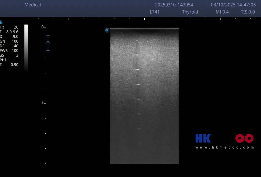
1.Vertical target group: It consists of 19 target lines, and the center distance between each adjacent pair of lines is 10mm.
2. Tumor-like phantom: The center depth is set at 30, 70, and 110 mm from the upper edge of the phantom, presenting a cylindrical shape with a diameter of 9 mm. The axis of the cylinder is parallel to the treatment line.
3. Mimic cyst: The center depth of the mimic cyst is located at 30 and 70 mm from the upper edge of the phantom, presenting a cylindrical shape with a diameter of 6 mm. The center depth of the mimic cyst is at 110 mm, also presenting a cylindrical shape with a diameter of 9 mm. The axis of the cylinder is parallel to the treatment line.
Note: Due to the exposed nature of the body model material, in order to maintain the reusability of the body model, during storage and use, please ensure that an additional layer of ultrasonic maintenance solution is placed on top of the internal tissue-like material.
The internal target types, sizes, and positions of this puncture body model can be customized according to your requirements. Additionally, other types of ultrasonic tissue-like target lesions can also be embedded inside the body model as per your specifications.
The puncture training mannequin set is a custom-made product. It is equipped with materials that simulate human tissues and is used for ultrasound imaging puncture training. The model is named KS105FC-1. It is filled with ultrasound-simulated human tissue materials. Inside the mannequin, there is a row of depth linear target lines arranged along the depth direction. On both sides of the target lines, cylindrical target markers such as tumors and cysts are buried. The depths of the simulated tumors are 30mm, 70mm, and 110mm, with a diameter of 9mm. The axes of the lesion columns are parallel to the target lines. The center depth of the 6mm-diameter simulated tumor is 30mm and 70mm, and the depth of the 9mm-diameter simulated cyst is 110mm. This mannequin is used to complement ultrasound-guided puncture training and demonstrations. It can be used for multiple punctures. The puncture holes can be closed after the puncture needle is removed, and there will be no puncture marks in the ultrasound image.
Basic Structure :
1. The outer shell is made of acrylic glass.
2. The overall dimensions are 220mm × 220mm × 75mm.
3. Built-in uniform human-like material.
5. Total weight: approximately 5 kilograms.
Technical Characteristics:
According to the national standard GB10152—2009, the technical parameters of the ultrasonic body model for training needs are as follows:
The inner material (imitating human body material) has a sound velocity of: (1540 ± 10) m/s (23℃ ± 3℃)
Attenuation coefficient slope: 0.50 ± 0.05 dB/cm/MHz (23 ± 3℃)
Dynamic elastic modulus E: 36 ± 2 KPa (at 23℃)
Built-in:
1.Vertical target group: It consists of 19 target lines, and the center distance between each adjacent pair of lines is 10mm.
2. Tumor-like phantom: The center depth is set at 30, 70, and 110 mm from the upper edge of the phantom, presenting a cylindrical shape with a diameter of 9 mm. The axis of the cylinder is parallel to the treatment line.
3. Mimic cyst: The center depth of the mimic cyst is located at 30 and 70 mm from the upper edge of the phantom, presenting a cylindrical shape with a diameter of 6 mm. The center depth of the mimic cyst is at 110 mm, also presenting a cylindrical shape with a diameter of 9 mm. The axis of the cylinder is parallel to the treatment line.
Note: Due to the exposed nature of the body model material, in order to maintain the reusability of the body model, during storage and use, please ensure that an additional layer of ultrasonic maintenance solution is placed on top of the internal tissue-like material.
The internal target types, sizes, and positions of this puncture body model can be customized according to your requirements. Additionally, other types of ultrasonic tissue-like target lesions can also be embedded inside the body model as per your specifications.
SAG: KS105FC-1 Puncture Phantom,Ultrasound Guided Biopsy,Training Phantom,Interventional Ultrasound,Medical Simulation,Biopsy Training,QC Phantom,Ultrasound Phantom,Tissue Mimicking,Clinical Skills